Team:Toulouse-INSA-UPS/Experiments

EXPERIMENTS


Here, you will find all the necessary information about the wetlab materials and methods we used in the course of our project.

Escherichia coli vectors
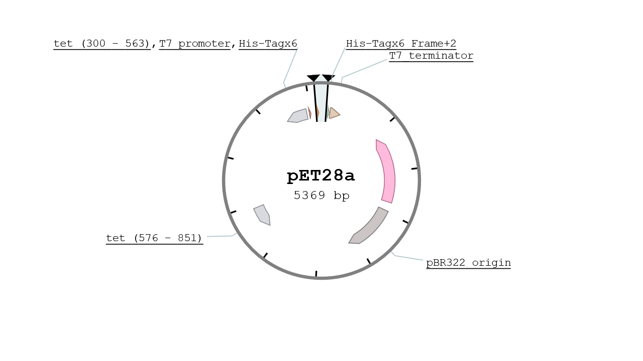
pET28

The pET28 vector contains two His Tags in its MCS and is specified for protein expression at high yield. It gives the strain a resistance to Kanamycin and its replication origin is the pBR322 which is a mid - low copy number plasmid (~10). The vector used was a kind gift from IBCG library.

A generic square placeholder image with rounded corners in a figure.
Figure 1. Map of the pET28 with the MCS Highligthed

pETDuet-1

The pETDuet-1 is, like the pET28, specialized in high yield protein production. Its main difference is that it contains two MCS and can therefore express two proteins at the same time under the same expression conditions. It bears ampicilin resistance and is a mid - low copy number plasmid. The plasmid used was a gift from IBCG library.

A generic square placeholder image with rounded corners in a figure.
Figure 2. Map of the pETDuet-1

Pichia pastoris vectors

pPICZ alpha

The pPICZalpha is an integrative plasmid for Pichia pastoris protein expression and suitable for E. coli replication. Its MCS is suitable for protein expession using the methanol inductible AOX1 Pomoter (PAOX1) and offering the alpha factor secretion signal for protein purification from culture supernatant. It contains a His tag and a Myc Tag for affinity purification stategies.

A generic square placeholder image with rounded corners in a figure.
Figure 3. Map of the pPICZ alpha

pGAPZalpha

The pGAPZalpha is roughly the same as the pPICZalpha with a different promoter that is the GAP Promoter, a glucose inductible promoter.

A generic square placeholder image with rounded corners in a figure.
Figure 4. Map of the pGAPZ alpha

Primer used

  • Cerberus Forward : TAAGAAGGAGATATACCATGGCGGAAGCGGGTATCACC
  • Cerberus Reverse : CTCGAGTGCGGCCGCAAGCTTCGGATCGTCCTATGATGGAGG
  • Sirius Forward: TAAGAAGGAGATATACCATGAATGCTACGCCAACTAAGGGTGC
  • Sirius Reverse: CTCGAGTGCGGCCGCAAGCTTAGCACCGGTGGAGTGACG
  • BirA Forward: AAGGAGATATACATATGAAGGATAACACCGTGCCACTGA
  • BirA Reverse: CTTTACCAGACTCGATTATTTTTCTGCACTACGCAGGGA
  • BFP Forward: ACCACAGCCAGGATCCTATGAGCGAACTGATCAAAGAGAACA
  • BFP Reverse: ATGCGGCCGCAAGCTTCTCATGCCATTCAATTTTCTGTGCT
  • RFP Forward: AGGAGATATACCATGGCTTCCTCCGAAGACGTTATCAAAG
  • RFP Reverse: GTGCGGCCGCAAGCTTAGCACCGGTGGAGTGACG
  • Scygonadin Forward: GGCTGAAGCTGAATTCGGCCAGGCACTCAACAAAC
  • Scygonadin Reverse: TGGGCCACGTGAATTCTCACTCATGCCATTCAATTTTCTG

Escherichia coli strains

Plasmid amplification strains
  • Stellar F-, endA1, supE44, thi-1, recA1, relA1, gyrA96, phoA, Φ80d lacZΔ M15, Δ(lacZYA-argF) U169, Δ(mrr-hsdRMS-mcrBC), ΔmcrA, λ-
  • Top10 F- mcrA Δ(mrr-hsdRMS-mcrBC) φ80lacZΔM15 ΔlacX74 nupG recA1 araD139 Δ(ara-leu)7697 galE15 galK16 rpsL(StrR) endA1 λ-
Protein production strains
  • BL21(DE3) B F-ompT gal dcm lon hsdSB(rB-mB-) λ(DE3[lacI lacUV5-T7p07 ind1 sam7 nin5])[malB+]K-12S)
  • Tuner F- ompT hsdSB (rB- mB-) gal dcm lacY1(DE3)

Pichia pastoris strains

For protein production we used GS200(ΔARG4, ΔHIS4) Strain for Cerberus production and X33(wt) for Scygonadin

Gluconacetobacter hansenii strain ATCC 53582

Click reagants

  • DBCO-Biotin, Sigma CAS : 1255942-07-4
  • DBCO-Fluorescein, Jena Bioscience Cat. No. : CLK-051-1
  • DBCO-Magnetic beads, Jena Bioscience Cat. No. : CLK-1037-1
  • Fluorescein-Azide, Jena Bioscience Cat. No. : CLK-80101-5
  • 4-L-azidophenylanaline, Sigma Ref. : 06162

Graphene Functionalization

  • Graphene, Sigma Ref. : 900561
  • 4-Ethynylaniline, Sigma Ref. : 481122
  • Isopentyl Nitrite, Sigma Ref. : 150495
  • N,N-Dimethylformamide, Sigma Ref. : 227056

Protein purification

  • Cobalt Affinity Gel, Sigma Ref. : H8162
  • Protease inhibitor cocktail tablet, Sigma Ref. : S8830

Cellulose Binding assay

  • Avicel, Sigma Ref. : 11365

Media


To prepare each medium, autoclave 20 minutes at 121°C.

LB Medium


Component Concentration (g/L)
Tryptone 10
NaCl 10
Yeast extract 5
**Agar 15

** Only for solid medium.

Nb: For culture with zeocin as an antibiotic, the NaCl concentration has to be 4g/L.

HS Medium


Component Concentration (g/L)
Glucose 20g
Yeast extract 5g
Peptone 5g
Na2HPO4 2.7g
Citric acid 1.5g
**Agar 15g

** Only for solid medium.

NB: For a 1 liter media, add 500ml of dionized water to glucose in one bottle and 500ml of dionized water to the rest in a second bottle. In incompletely distilled water, glucose will form a solid mass, so stir vigorously immediately after adding water. Autoclave both bottles to sterilize media and pour glucose solution in sterile conditions (next to a Bunsen burner or in a flow hood) into the second bottle.
Autoclaving glucose separately from amino acids avoids Maillard reaction, which can result in the formation of toxic byproducts in the media.

YPD Medium


Component Concentration (g/L)
Peptone 20
Glucose 20
Yeast extract 10
**Agar 15

** Only for solid medium

NB: Autoclave the glucose solution separately from the rest.
Autoclaving glucose separately from amino acids avoids Maillard reaction, which can result in the formation of toxic byproducts in the media.

TB medium


Component Concentration (g/L)
Tryptone 12
Yeast extract 24
Glycerol 4mL

For solid media, add 15g of agarose per liter.
Autoclave : 20min at 121°C

Minimal Dextrose Medium (MD)


Medium for selection of recombinant Arg+ clones For 1 L MD agar plates you need:

  • Autoclave 2 g dextrose and 15 g agar
  • L-Histidine (0.04% w/v)
  • Add 100 mL sterile 10X YNB stock solution
  • 2 mL sterile 500x biotin stock solution

Yeast Nitrogen Base (YNB) 10X stock solution:

  • Dissolve 69 g YNB (without aminoacids; with ammonium sulfat) in 500 mL bidest water and filter sterilize.
  • Store at 4 °C.
  • Will last for one year.

Biotin 500X stock solution:

  • Dissolve 20 mg biotin in 100 mL of 0.05 M NaOH solution and filter sterilize.
  • Store at 4 °C.
  • Durable for one year.

BMMY medium


BMMY Buffered Methal-complex medium

Component Concentration (g/L)
Peptone 2%
Yeast extract 1%
YNB 1.34%
Biotin 0.4 µg/mL
Methanol 0.5%
Potassium phosphate pH 6 100mM

Storage conditions : 4°C

BMGY medium


BMGY Buffered Glycerol-complex medium

Component Concentration (g/L)
Peptone 2%
Yeast extract 1%
YNB 1.34%
Biotin 0.4 µg/mL
Glycerol 1%
Potassium phosphate pH 6 100mM

Storage conditions : 4°C

Buffer


Materials


  • pH meter
  • NaOH solution
  • HCl 90%
  • kOH
  • Acetic acid

Buffer for transformation


YETM pH 7.5


Component Concentration (g/L)
Tryptone 20
MgSO4-7H20 10
Yeast extract 5
**Agar 15

** Only for solid medium

Adjust to pH to 7.5 with kOH.

TFB1 pH 5.8


Component Concentration (g/L)
C2H302K (30 mM) 2.95
RbCl (100 mM) 12.1
CaCl2-2H2O (10 mM) 1.45
MnCl2-4H2O (50 mM) 9.9
Glycerol (100 %wt/vol) 22.5

Adjust to pH 5.8 with 0.2M acetic acid. Add dionized water up to 1L. Filter sterilize. Store refrigerated at 4°C.

TFB2 pH 6.5


Component Concentration (g/L)
MOPs (10 mM) 2.1
CaCl2-2H2O (75 mM) 11.05
RbCl (10 mM) 1.2
Glycerol (100 %wt/vol) 22.5

Adjust to pH 6.5 with kOH. Add dionized water to 1L. Filter sterilize. Store refrigerated at 4°C.

Guanidine HCl 6M pH 7.5


Component Quantity
Guanidine 286.59g
deionized water 500 L

To regulate the pH use NaOH of HCl 90%.
Store at room temperature.

Culture conditions


Escherichia coli

  • 37°C
  • 160 rpm (for liquid media)

Pichia pastoris

  • 37 °C
  • 160 rpm (for liquid media)

Gluconacetobacter hansenii

  • 30°C
  • Standing
  • HS medium

Cloning


1 . Insert preparation


PCR ClonAMP®

Component Quantity
Premix ClonAMP® PCR 25µl
Primer 1 1.25µl
Primer 2 1.25µl
DNA (0.5 ng/µl) 4µl
Deionized water MQ 18.5µl

PCR ClonAMP program :

  • Cycle 1 : - 98 °C 2min
  • Cycle 2 : - 98°C 10s
    - 60°C 5s
    - 72°C 5s/kb
  • Cycle 3 : - 72°C 2 min

2 . Vector preparation


2 . 1 . Miniprep and Midiprep
  • Miniprep
This protocol is extracted from the GenElute™ Plasmid Miniprep Kit from Sigma Aldricht.

  1. Harvest cells
    - High-copy plasmid : 1-3 ml
    - Low-copy plasmid : 1-5 ml
    Pellet 1-5ml of an overnight recombinant E. coli culture by centrifuging at 4,500rpm for 4min. Discard the supernatant.
  2. Resuspend cells
    Completely resuspend the bacterial pellet with 200 µl of the Resuspension Solution. Vortex or pipette up and down to thoroughly mix the cells until homogeneous.
  3. Lyse cells
    Lyse the resuspended cells by adding 200 µl of the lysis Solution. Immediately mix the contents by gentle inversion (6-8 times) until the mixture becomes clear and viscous.
    NB : Do not allow the lysis reaction to exceed 5 minutes.
  4. Neutralize
    Add 350 µl of neutralize solution. Gently invert the tube 4-6 times.
    Pellet the cell debris by centrifuging at 12,000 x g during 10 minutes.
  5. Prepare column
    Insert a GenElute Miniprep Binding Column into a provided microcentrifuge tube.
    Add 500 µl of the Column Preparation Solution to each miniprep column and centrifuge 12,000 x g during 1min.
  6. Load cleared lysate
    Transfer the cleared lysate from step 4 to the column prepared in step 5 and centrifuge at 12,000 x g for 1 min. Discard the flow-through liquid.
    Centrifuge again at 12,000 x g for 1 min.
  7. Wash column
    Add 750 µl of the diluted Wash Solution to the column.
    Centrifuge at 12,000 x g for 1 min.
    Discard the flow-through liquid.
    Centrifuge again at 12,000 x g for 1 min.
  8. Elute DNA
    Transfer the column to a fresh collection tube. Add 50 µl of deionized water (MiliQ).
    Wait 2-3 minutes.
    Centrifuge 1 minute at 12,000 x g.
    The eluate is ready for immediate use of storage at -20°C.
  • Midiprep

This protocol is extracted from the QIAfilter™ Plasmid Midi Kit (25) from QIAGEN.

High-copy plasmid : 25 ml
Low-copy plasmid : 50-100 ml

  1. Harvest bacterial culture after 12-16 h of growth by centriuging at 6000 x g for 15 min at 4°C.
  2. Completely resuspend in 4 ml of Buffer P1
  3. Add 4 ml Buffer P2 by inverting the sealed tube 4-6 times and incubate at room temperature for up to 5 min. No more than 5 min !!!
  4. During the incubation, screw the cap onto the outlet nozzle of the QIAfilter Cartridge, and place it in a convenient tube or a QIArack
  5. Add 4 ml prechilled Buffer P3 to the lysate, and mix the solution until it is completely colorless.
  6. Pour the lysate into the barrel of the QIAfilter Cartridge. Incubate at room temperature for up to 10min. DO NOT INSERT THE PLUNGER !
  7. Equilibrate the QIAGEN-tip by applying 4 ml Buffer QBT, and allow the column to empty by gravity flow.
  8. Remove the cap from the QIAfilter Cartridge outlet nozzle. Gently insert the plunger into the QIAfilter Cartridge, and filter the cell lysate into the equilibrated QIAGEN-tip. Allow the lysate to enter the resin by gravity flow.
  9. Wash the QIAGEN-tip with 20 ml of Buffer QC
  10. Elute the DNA with 5ml Buffer QF.
  11. Precipitate DNA by adding 3.5 ml isopropanol mix and centrifuge at 4000 rpm for 30min at 4°C. Carefully decant the supernatant.
  12. Wash the DNA pelletwith 5 ml ethanol 70 % and centrifuge at 4000 rpm for 10 min at 4°C. Carefully decant the supernant.
  13. Air-dry the pellet for 20 min and redissolve DNA in 150 µl miliQ water
2 . 3 . Preparative digest gel extraction

Component Quantity
DNA plasmid 5-10µg
Cutsmart® buffer 5µl
Enzyme** 1µl
Deionized water MQ up to 50µl

​​​​​​​** If you have 2 enzymes 0.5µl of each ones.

3 . Gel extraction purification


This protocol is extracted from the NucleoSpin® kit. We would extract both vector and insert with this protocol.

  1. Excise DNA fragment and weight it.
  2. Solubilize gel slice : for each 100 mg of agarose gel, add 200 µl of Buffer NTI. Incubate sample for 10 min at 50°C, mix by inverting the tube every 2-3 min.
  3. Bind DNA : place a column (provided in the kit) into a collection tube (2 ml, also provided) and load up to 700µl sample. Centrifuge for 30s at 11,000g. Discard flow-through and place the column back into the collection tube.
  4. Wash silica membrane : add 700µl Buffer NT3 to the column. Centrifuge for 30s at 11,000g. Discard flow-through and place the column back into the collection tube. Repeat this step one more time.
  5. Dry silica membrane : centrifuge 1 min at 11,000g to remove Buffer NT3 completely.
  6. Elute DNA : place the column into a new 1.5ml Eppendorf. Add 20 µl of H2O MQ and incubate at room temperature (18 - 25°C) for 1 min. Centrifuge for 1 min at 11,000g.

4 . InFusion cloning


4 . 1 . Cloning

Materials

  1. Insert : PCR Fragment purified (ClonAMP®)
  2. Vector : Linearized plasmid purified (Cutsmart®)
  3. In-Fusion® cloning kit Takara
  4. PCR thermocycler

Protocol

Rxn Component Cloning Rxn
Purified PCR fragment 10 - 200 ng*
Linearized plasmid 50 - 200 ng*
5X Fusion HD enzyme premix 2µl
Deionized up to 10 µl
4 . 2. Transformation into Stellar® competent cells

Transformation


1 . Escherichia coli


1 . 1 . Preparation of competent cells : BL21(DE3)


Protocol

  1. Streak cells from frozen stock onto LB plate. Incubate O/N at 37°C.
  2. Pick a single fresh colony to inoculate 5 mL of LB medium. Grow O/N at 37°C.
    Do not vortex cells at any time after this point in the procedure.
  3. Dilute 1 mL of culture into 50 mL LB medium prewarmed to 37°C. Grow at 37°C for 2 hours with agitation.
    Volumes can be scaled up 5X and all of the 5 mL overnight culture can be used.
  4. Transfer culture to sterile 50 mL tube. Chill on ice 10-15 minutes.
  5. Centrifuge for 10 mintutes at 2000 rpm at 4°C. Immediately aspirate off all the supernatant.
    Do not allow cells to warm above 4°C at any time in this procedure.
  6. Resuspend cells in 10 mL of ice-cold TFB1 with gentle re-pipetting. Use chilled glass of plastic pipette.
  7. Incubate cells on ice for 5 minutes.
  8. Repeat step 8.
  9. Resuspend cells in 2 mL of ice-cold TFB2 with gentle re-pipetting. Use micropipet tip (plastic).
  10. Incubate cells on ice for 15 minutes. Cells may be used for transformation or frozen.
    To freeze: aliquot cells 100 μL volumes into prechilled 0.5 mL microcentrifuge tubes (on ice). Freeze
    immediately on dry ice. Stire cells frozen at -80°C.

1 . 2 . Transformation of Competent Cells


Protocol

  1. If starting with frozen competent cells, warm tube/cells by gently twirling between your fingers until just thawed
    (i.e., at ~0°C). Then, immediately place on ice for about 5 minutes.
  2. Set up transformation as follows:
    Add to 15 mL plastic round bottom tube on ice:
    0-9 μL TE (Tris 10mM + EDTA 1mM)
    1-10 μL DNA (10-100 ng)
    10 μL final volume --> /!\ 10% max of the cell competent volume
  3. Add 100 μL of competent cells and mix by gentle repipetting.
    This method can be scaled down 2- to 4-fold. The maximum volume of DNA should be ~1/10 volume of cells
    and the maximum mass should be <= 100 ng of DNA for 100 μL of cells.
  4. Incubate cells on ice for 20-30 minutes.
  5. Heat shock the cells exactly 90 seconds at 42°C.
  6. Return cells on ice 2 minutes.
  7. Add 1 mL of LB medium. Incubate at 37°C for 45-60 minutes with slow gentle shaking.
    For blue/white color selection, spread IPTG and X-gal on plates now and hold at 37°C until use.
  8. Plate 0.1 - 0.2 mL of transformed cells on LB-plate containing the appropriate antibiotic.
  9. Incubate overnight at 37°C. Place at 4°C to store.

Testing competent cells

  1. Transform 100 μL of cells with 1 μL (10 pg) of pUC19 monomer (0.01 μg/μL).
  2. Plate 0.25 mL of transformation mixture. Incubate overnight at 37°C.
  3. Count CFU and calculate efficiency. Efficiency of colonies per μg of colonies 4 * 105. You should
    obtain 1-5 X 10⁷/μgDNA from competent cells after one freeze-thaw cycle.

2 . Pichia pastoris


2 . 1 . Electroporation


2 . 1 . 1 . Competent cells preparation

Materials
- YPD/0.02M HEPES buffer
- 1.0M DTT (Dithiothreitol)
- 1.0M Sorbitol
- 1.5ml tubes
- Sterile water (1.250l)
- Centrifuge (with temperature control)

Protocol

  1. Grow 500ml cells to DO 1.3 to 1.5 at 600 nm and keep in ice for the duration of the procedure)
  2. Centrifuge cells for 5 min (all centrifugation steps were at 4000g at 8°C)
  3. Add 100ml YPD/0.02M HEPES
  4. Add 2.5ml 1.0M DTT dropwise
  5. Incubate for 15 min at 30°C with shaking (150 rpm)
  6. Add water to 500ml
  7. Centrifuge 5 min
  8. Remove the supernatant and resuspend in 500 ml water
  9. Centrifuge 5 min
  10. Remove the supernatant and resuspend in 250 ml water
  11. Centrifuge 5 min
  12. Remove the supernatant and resuspend in 20 ml sorbitol (1M)
  13. Centrifuge 5 min
  14. Remove the supernatant and resuspend in 1 ml sorbitol (1M)
  15. Aliquot into 40 µl in individual 1.5 ml tubes
  16. Place in -80°C freezer until needed
2 . 1 . 2 Electroporation Pichia pastoris

Materials:
 - Ice
 - Linearized plasmid
 - Competent cells
 - Electroporation apparatus
 - 1.0M Sorbitol
 - YPD
 - Plates with gradient of Zeocin
​​​​​
Protocol

  1. Mix 5μL (50–100 ng) of linearized plasmid DNA with 40 μL of competent cells in an electroporation cuvette. Tap the cuvette on the table to take away the air bubbles.
  2. Incubate for 2 min on ice.
  3. Have two pipettes ready before electroporation: one with 0.5 ml of 1.0 M Sorbitol and one with 0.5 ml YPD.
  4. Pulse 1500V, 25μF, 200Ω (program Sc2). You should have a Ꞇ between 4 and 5 ms.
  5. Resuspend immediately samples in 0.5 mL 1.0 M sorbitol and 0.5 mL YPD, incubate in a 30°C shaker for 1h 30, and then plate on media containing increasing concentrations of zeocin for the selection of multicop.

It can take around one week to have one colony on plate.

2 . 2 . Heat shock Pichia pastoris


2 . 2 . 1 . Cell Preparation

Protocols

  1. Grow 10 mL cells to desired A600
  2. Centrifuge cells at 4000g at 4°C for 5 min
  3. Resuspend in 10 mL BEDS solution
  4. Centrifuge cells for 5 min
  5. Resuspend cells in 1 mL BEDS
  6. Aliquot into individual 1.5 mL tubes
  7. Place in −80°C freezer until needed

BEDS solution is composed of 10 mM bicine-NaOH, pH 8.3, 3% (v/v) ethylene glycol, 5% (v/v) dimethyl sulfoxide (DMSO), and 1 M sorbitol.

2 . 2 . 2 Transformation
  1. Mix DNA with cells (50 µl cells, 5-8 µl DNA)
  2. Add 1.4 ml 40% polyethylene glycol (PEG), 200 mM bicine, pH 8.3
  3. Incubate for 60 min at 30°C with agitation at 130 rpm
  4. Heat shock at 42°C for 10 min (optional recovery for 1–3 h)
  5. Pellet cells for 5 min
  6. Resuspend cells with 1ml of 150 mM NaCl, 10 mM bicine, pH 8.3
  7. Repeat steps 5 and 6
  8. Plate

3 . Transformation for InFusion

Stellar® competent


Protocol

  1. Thaw Stellar® competent cells on ice just before se. After thawing, mix gently to ensure even distribution, and then move 50µl of competent cells into a 14ml round bottom tube (falcon tube). Do not vortex.
  2. Add 2.5 µl of the in-fusion reaction mixture to the competent cells.
  3. Place the tubes on ice for 30 min.
  4. Heat shock the cells for exactly 45sec at 42°C.
  5. Place the tubes on ice for 1-2min.
  6. Add soc medium to bring the final volume to 500µl. SOC medium should be warmed at 37°C before using.
  7. Incubate 1hour at 37°C.
  8. Place 1/100 -1/5 of each transformation reaction into separate tubes and bring the volume to 100µl with SOC medium. Spread each diluted transformation reaction on separate LB plate containing an antibiotic.
  9. Centrifuge the reminder of each transformation reaction at 6 000 rpm for 5min. Discard the supernatant and resuspend each pellet in 100µl freash SOC medium. Spread each sample on a separate LB plate containing the appropriate antibiotic. Incubate of the plates overnight at 37°C.
  10. The next day, pick individual isolated colonies from each experimental plate. Isolate plasmid DNA using standard method of your choice (e.g miniprep). To determine the present of an insert, analyze the DNA by restriction digestion or PCR screening.

Protein production


1. Protein production - Escherichia coli


Material

  • LB 10 g/L NaCl (800 ml)
  • Your favourite antibiotic
  • LB agar plates containing your favourite antibiotic
  • IPTG 1M (~1ml)
  • Talon Buffer 1x 10mM Imidazole

Protocol

  • Day one :
    • Transform your plasmid in E. coli BL21(DE3) competent cells plate on LB Agar + Your favourite antibiotic and incubate O/N at 37°C
  • Day two :
    • Pick 3 colonies and seed then in 9ml LB + Your favourite antibiotic. Incubate O/N at 37°C under 130 rpm shaking.
    • Autoclave 800ml LB 10 g/L NaCl in a 2L baffled flask and preheat it at 37°C O/N.
  • Day three:
    • Take a sample of the 800ml culture and measure OD600 as blank.
    • Take 1ml of preculture and measure OD600, seed the preculture in the 800ml LB and take OD.
    • Calculate OD600 assuming that it doubles every 20 minutes and check the OD until it reaches 0.5 ~0.6. Once the exponontial phase is reached, induce with 800µl of 1M IPTG stock solution and incubate at 37°C for 4h or O/N at 16°C.
    • Pellet cells by centrifugate at 5000 x g for 10 minutes and resuspend in 10ml TB 1x 10mM Imidazole if growth was performed at 37°C of 10ml of O/N.
    • Freeze cells for later purification or wait at least 30 minutes to continue the protocol.
    • Sonication : Run on cycle of 1 minute and one of 30 seconds, both at 25 of intensity.
    • Pellet cells by centrifugate at 60,000 x g for 30 minutes and withdraw the supernatant that is the cell free extract.
    • Then continue to the protein purification protocol.

2. Protein production - Pichia pastoris


Expression can be done in either YPD medium (1% yeast extract, 2% peptone, 2% glucose) or Yeast Nitrogen Base with 0.5% glucose.

  1. Using a single colony, inoculate 10 ml of YPD. Grow at 28–30°C in a shaking incubator (250–300 rpm) O/N.
  2. Use 0.1 ml of the overnight culture to inoculate 50 ml of YPD in a 250 ml flask. Grow at 28–30°C in a shaking incubator (250–300 rpm).
  3. At each of the times indicated below, transfer 1 ml of the expression culture into a 1.5-ml microcentrifuge tube. These samples will be used to analyze expression levels and determine the optimal time to harvest. Centrifuge at maximum speed in a tabletop microcentrifuge for 2–3 minutes at room temperature.
    Time points (hours): 0,24 (1 day), 48 (2 days), 72 (3 days) 96 (4 days)
  4. For secreted expression, transfer the supernatant to a separate tube. Store the supernatant and the cell pellets at –80°C until ready to assay. Freeze quickly in liquid N2 or a dry ice/ethanol bath.
  5. For intracellular expression, decant the supernatant and store just the cell pellets at –80°C until ready to assay. Freeze quickly in liquid nitrogen or a dry ice/ethanol bath.

Protein purification


1. Purification for His-tagged protein


1 . 1 .IMAC column purification

The volumes will be expressed as CV (Column Volume) which refers to the volume of resin deposited.

Materials

  • TB 10mM Imidazole
  • TB 40mM Imidazole
  • TB 100mM Imidazole
  • TB 300mM Imidazole
  • IMAC column
  • His-Select Cobalt Affinity Gel

Protocol

  1. Equilibrate column with 10 CV TB 10mM Imidazole
  2. Wash with at least 2 CV 2 or 3 times
  3. Elute with 2 CV of :
    • 40mM one time
    • 100mM 2 times
    • 300mM 1 time
  4. Analyse different fractions by SDS PAGE.

The protocol must be adapted depending on the protein studied as the production yields will be protein dependant.

Cleaning His-Select Cobalt Affinity Gel

  1. Wash the affinity gel with 2 column volumes of deionized water
  2. Clean with 5 column volumes of 6M guanidine HCl, pH 7.5. The flow rate should be no more than 5 column volumes per hour.
  3. Remove the guanidine HCl solution by washing with 2-3 column volumes of deionized water.
  4. For immediate use, re-equilibrate the affinity gel with 2-3 column volumes of Equilibration Buffer. If the gel is to be stored, wash with 1-2 column volumes of 30% ethanol and store at 2-8°C.

2 . Purification for CBM protein


2 . 1 . RAC preparation


Materials

  • Cellulose
  • H2PO3
  • Na2CO3 2M
  • Deionized water
  • Ice

Protocol

  1. Weigth about 0.2 g of Cellulose, put it in a 50ml Falcon tube and add 0.6 ml of H20 to wet it.
  2. Add 10ml of ice cold H2PO3 and vortex
    incubate 1h on ice and vortex envery 20 minutes
  3. Add 40 ml of ice cold H2O and vortex every 10ml
    • before adding the last 10ml vortex hard
  4. Centrifugate at 10,000 x g for 20 minutes and discart supernatant.
  5. Wash with 45ml of H20 and 0.5ml of Na2CO3 2M
    • Repeat 4 times
  6. Resuspend in 20 ml of Water to a final concentration of 10mg/ml

2 . 2 . CBM protein purification : Cellulose affinity


Material

  • Cell Free Extract
  • Regenerated Amorphous Cellulose
  • Ethylene Glycol
  • RAC previously prepared

Method

  • Add 5ml of RAC (10g/L) to the Cell Free Extract, volumes can be scaled up depending on the production yield required. Mix by inverting and incubate 30 minutes at room temperature.
  • Centrifugate 4 500 x g for 20 minutes. Collect supernatant as Flow Through fraction. Resuspend pellet with 5 RAC Volume of 50mM Tris HCl pH 8.
  • Centrifugate at 4 500 x g for 20 minutes and discart supernatant, repeat twice
  • Resuspend pellet in 2 RAC Volumes of 100% Ethylene Glycol and let sit for 15 minutes.
  • Centrifugate at 4 500 x g for 20 minutes, Supernatant can be conserved at 4°C or -20°C depending on the usage required.
  • Analyse by SDS PAGE.

3 .Protein concentration using vivaspin column


Materials

  • Vivaspin column

Protocol
Add your favourite purified protein (Dont go over 20ml) and equilibrate with and empty tube willed with water (DO NOT DISCART AFTER) and centrifugate for 5 minutes at 4500 x g. Observe the eluted volume and adapt the time for the desired final volume (this is protein dependant).

4 . Desalting protein using PD-10 column


  1. Remove the top cap and cut the sealed end of the column
  • let the storage solution elute
  • Fill the column with the equilibration buffer (the one you will want your protein to be recovered in
    • Repeat 4 times
    • Discart the Flow Through
    • (Around 25ml should be used in total)
  1. Add max 2.5 ml of the sample to the column and let it enter completely
  • If sample volume is less than 2.5ml add equilibration buffer to complete AFTER the sample has entered the column completely
  • Discart the flow through
  1. Place a collection tube and elute with 3.5ml buffer.
  • If larger volume are needed to be concentrated repeat initial Equilibration step before adding another one.

Cellulose pull down assay


Material

  • Regenerated Amorphous Cellulose 10 mg/ml
  • 50mM Tris HCl pH 8
  • Protein sample (concentration about 5mg/ml)
  • Ethylene glycol
  • RAC previously prepared

Method

  • Prepare solutions following these amounts :
    • 75µl Tris HCl pH 8
    • 100µl RAC
    • 4µl protein sample (prepare negative control by replacing it with water)
    • 21µl H20 mQ (can aslo be replaced with protein for saturating the RAC)
  • Incubate at room temperature for 30 minutes
  • Centrifugate at >10,000 x g for 10 minutes
    • Collect supernatant fraction
  • Wash with 5 x RAC Volume of Tris HCl
    • Centrifugate at >10,000 x g for 10 minutes
  • Eluate protein with 2 x RAC Volume of Ethylene glycol
  • Analyse different fractions by SDS PAGE.

Strain Promoted Alkyne Azide Cycloaddition

Method

Add in 100μL a ratio of 1:10 of Azide:DBCO, cover with aluminium foil and incubate O/N under shaking