Kristinazu (Talk | contribs) |
|||
| Line 46: | Line 46: | ||
</section> | </section> | ||
<section class="design_subsections"> | <section class="design_subsections"> | ||
| − | + | <h1 id="Ribosome_modifications">Ribosome modifications</h1> | |
| − | + | <div class="third_level_links"> | |
| − | + | <a href="#Liposomes">Liposomes</a> | |
| − | + | <a href="#Ribosome_modifications">Ribosome modifications</a> | |
| − | + | <a href="#BAM complex">BAM complex</a> | |
| − | + | <a href="#RNA_Thermoswitches">RNA Thermoswitches</a> | |
| − | + | <a href="#Mistic_fusion_protein">Mistic fusion protein</a> | |
| − | + | <a href="#Surface_display_system">Surface display system</a> | |
| − | + | </div> | |
| − | + | <div> | |
| − | + | <h1> | |
| − | + | Background | |
| − | + | </h1> | |
| − | + | <p></p> | |
| − | + | <p> | |
| − | + | Insertion of many membrane proteins in prokaryotes as well as the endoplasmic reticulum of eukaryotes is facilitated by various translocons. These complexes interact with the ribosome during protein synthesis and direct the newly forming peptide into the translocon pore, or directly into the membrane. However, for the translocon to function properly, it requires many auxiliary components (signalling sequence, chaperones, insertion mechanism), which would be far too many for our system to remain stable. Since we could not simply transfer the entire translocation system into SynDrop, we hypothesized that some of its functionality could be emulated in different ways. | |
| − | + | </p> | |
| − | + | <p> | |
| − | + | <h2> | |
| − | + | Nickel-chelating lipids and polyhistidine tags as potential solution | |
| − | </div> | + | </h2> |
| + | </p> | ||
| + | <p> | ||
| + | Specifically to localize the ribosomes near the membrane and reduce the exposure of MP transmembrane domains to the aqueous environment, we eventually chose a method that has already been successfully used to attach other proteins to membranes - via a his-tag to nickel ions, chelated by specific lipids composing membrane<sup>1,2</sup>. Our reasoning for this was threefold. Firstly, this would have allowed ribosomes to be correctly positioned. If the ribosomes were localized near the membrane, but the peptide exit tunnel hadn’t been consistently pointed towards the membrane, the entire benefit of reducing MP aggregation would be lost. By being able to create his-tagged fusion proteins specifically of the ribosomal subunits localized near the exit tunnel, we guaranteed that the exit tunnel could never point away from the membrane. Secondly, general protein synthesis should be the least impeded, compared to alternatives, such as introducing a lipid anchor via an enzyme, or creating a fusion protein with a self inserting MP. These latter methods would completely immobilize the ribosome onto the membrane, removing degrees of freedom. With the polyhistidine-nickel interaction the ribosome would simply exhibit a much greater affinity towards the membrane, while still being able to detach and reattach as needed. Lastly, the simplicity for this method seemed to be the greatest, as only modified ribosomes as well as the incorporation of nickel-chelating lipids during liposome synthesis was needed - this was far more important than it can appear, because it was vital for our overall system to be as robust as possible. | ||
| + | </p> | ||
| + | <p> | ||
| + | <strong>Fig. 1</strong> Principle of ribosome attachment to the liposome membrane. The ribosome exit tunnel is localized near the membrane, resulting in transmembrane domains of newly synthesized peptides interacting with the membrane, reducing aggregation | ||
| + | </p> | ||
| + | <p></p> | ||
| + | <h1>Results</h1> | ||
| + | <p></p> | ||
| + | <p> | ||
| + | <h2>Designing the modifications</h2> | ||
| + | </p> | ||
| + | <p> | ||
| + | The basic procedure we designed to obtain our modified ribosomes is as follows: first, using CRISPR-Cas9, we modify the genome of E. coli (native strain MG1655) to incorporate his-tags onto the ribosomal subunits, as well as a Strep-tag onto the L12 subunit for ease of purification<sup>3</sup>. In literature a his-tag was described to aid purification when fused to subunit L12, however we exchanged it for a Strep tag, as it is also short in sequence and while being biologically does not interfere with the functionality of other his-tags. The subunits localized near the exit tunnel we chose as potential targets, in order of priority, were L24, L23, and L29. These subunits were chosen as they had free, unfolded C ends exposed to the surface at nearest proximity to the exit tunnel. These subunits are also not integral to the overall function of the ribosome, meaning that our modifications would have little to no impact on peptide synthesis<sup>4,5,6,7</sup>. After the E. coli genome is successfully modified, the ribosomes are purified with the help of the previously incorporated Strep tag, and the final product is used for IVTT reactions. | ||
| + | </p> | ||
| + | <p> | ||
| + | <h2> | ||
| + | Modification procedure | ||
| + | </h2> | ||
| + | </p> | ||
| + | <p> | ||
| + | CRISPR-Cas9 was utilized via pCas9 and pTargetF plasmids <sup>8</sup>. pCas9 constitutively expresses Cas9, which induces a double-strand DNA break at a specific target sequence, that is complementary to the guide RNA. The guide RNA sequences for our targets were introduced via reverse PCR into the pTargetF plasmid series, which then supplies it to Cas9. After the double stranded break occurs, the HDR (homology directed repair) mechanism is activated, which repairs the genome according to a supplied donor sequence. This process is highly efficient with the assistance λ-red proteins, expressed from pCas9. The donor sequence has ~300 bp length homology arms and the insertion sequence that can include either the His or Strep tag to be fused with the chosen ribosomal subunit C’-end, as well as an in frame selection marker (select antibiotic resistance genes). pCas9 expresses a gRNA targeting the ori of pTargetF, therefore the cells are automatically cured from pTargetF after each modification. Additionally, pCas9 has a temperature-sensitive ori, it is cured by growing the cells at 37<sup>o</sup>C. | ||
| + | </p> | ||
| + | <p> | ||
| + | <strong>Fig. 2</strong> Scheme of the genome modification process: | ||
| + | <ol> | ||
| + | <li> | ||
| + | 1. pCas9 is introduced into the target cells | ||
| + | </li> | ||
| + | <li> | ||
| + | 2. pTargetF and the according donor DNA are electroporated into the cells | ||
| + | </li> | ||
| + | <li> | ||
| + | 3. Cas9 and our custom gRNA form a complex that cuts the genome DNA at the target. The genome is repaired via HDR according to our donor DNA sequence - X1 (homology arm of target ribosomal subunit) with fused tag, X3 (antibiotic resistance gene) and X2 (second homology arm). | ||
| + | </li> | ||
| + | <li> | ||
| + | 4. pTargetF is cured and the process can be repeated with a new target. | ||
| + | </li> | ||
| + | </ol> | ||
| + | </p> | ||
| + | <p> | ||
| + | For multi-gene editing, we chose to supply the donor sequence as a linear DNA strand (PCR product). Due to financial reasons, to construct the donor DNA sequence we performed separate PCRs of the homology arms (from the E. coli genome), selection marker (antibiotic resistance genes from available plasmids) (Fig. 4). The oligomers had the his and strep tag sequences incorporated into them alongside 2 different restriction sites. In case the distance between the ribosomes and the membrane wall was too small for our system to be efficient, we also designed alternative variants the would feature the his-tags connected via a highly flexible two-glycine-four-serine linker (GGSSSS), which is a highly popular linker for artificial fusion proteins. | ||
| + | </p> | ||
| + | <p> | ||
| + | When the homology arm and marker gene pcr products are restricted and ligated, the end result is a single sequence with an antibiotic resistance gene flanked on either side by ~300 bp homology arms, in addition to the according tag fused to the selected subunit’s gene. The entire linear DNA product was amplified via PCR using primers that annealed to the ends of the strand. Large amounts of nonspecific product was present even after performing PCR of the whole ligated sequence, therefore gel purification was necessary after each amplification (Fig. 5). | ||
| + | </p> | ||
| + | <p> | ||
| + | <strong>Fig.3</strong> Example of a constructed donor sequence. The sequence of the selected tag is present in primer used for the PCR of the homology arm that encompasses the target subunit. As a result, the tag sequence is fused to the ribosomal subunit gene. | ||
| + | </p> | ||
| + | <p> | ||
| + | <strong>Fig. 4</strong> PCR of homology arms, and antibiotic resistance genes | ||
| + | </p> | ||
| + | <p> | ||
| + | <strong>Fig. 5</strong> Constructed donor DNA sequences. The L29 donor DNA was not further revisited due to time constraints | ||
| + | </p> | ||
| + | <p> | ||
| + | The genome modifications were then carried according to our protocol "kristina"(link). Although cPCR gave us mixed results, we could not verify any colonies that afterwards grew on our selected marker antibiotics, and thus could not continue our experiments with them. It appears most likely that the genome modifications were not entirely successful, due to the somewhat unstable nature of the ligated linear DNA used for the donor sequence. | ||
| + | </p> | ||
| + | <p></p> | ||
| + | <h1>Conclusion and Discussion</h1> | ||
| + | <p></p> | ||
| + | <p> | ||
| + | Although well described and planned in theory, our ribosome attachment system did not yield desired results. We hypothesize, that the underlying cause may include a flawed implementation of the CRISPR-Cas9 system: in our case we were forced to incorporate selection markers due to the functionality of the specific expression plasmids we used, which lead to the construction of unstable donor sequences. While this is unfortunate, we are still confident that our idea is worth further investigation: as we could not reproduce genome modifications on the subunit that has already been reportedly modified in a similar manner, it does not suggest that our primary concept is unworkable. Moving forward, we will most likely move to an alternative CRISPR-Cas9 expression system that would allow for optimized modifications, such as removing the requirement for a selection marker, which in turn would allow us to construct more robust donor sequences. Nonetheless, herein we provide a concept and initial tools for ribosome engineering. This demonstrates great potential in synthetic biology, especially in cell-free biomolecular systems like SynDrop. | ||
| + | </p> | ||
| + | <p></p> | ||
| + | <h2>References</h2> | ||
| + | <p> | ||
| + | <ol> | ||
| + | <li>1.Chikh, G., Li, W., Schutze-Redelmeier, M., Meunier, J. & Bally, M. Attaching histidine-tagged peptides and proteins to lipid-based carriers through use of metal-ion-chelating lipids. Biochimica et Biophysica Acta (BBA) - Biomembranes 1567, 204-212 (2002). | ||
| + | </li> | ||
| + | <li>2.Blanchette, C., Fischer, N., Corzett, M., Bench, G. & Hoeprich, P. Kinetic Analysis of His-Tagged Protein Binding to Nickel-Chelating Nanolipoprotein Particles. Bioconjugate Chemistry 21, 1321-1330 (2010). | ||
| + | </li> | ||
| + | <li>3.Ederth, J., Mandava, C., Dasgupta, S. & Sanyal, S. A single-step method for purification of active His-tagged ribosomes from a genetically engineered Escherichia coli. Nucleic Acids Research 37, e15-e15 (2008). | ||
| + | </li> | ||
| + | <li>4.Spillmann, S. & Nierhaus, K. The ribosomal protein L24 of Escherichia coli is an assembly protein. Journal of Biological Chemistry (1978). at http://www.jbc.org/content/253/19/7047.long | ||
| + | </li> | ||
| + | <li>5.GU, S. The signal recognition particle binds to protein L23 at the peptide exit of the Escherichia coli ribosome. RNA 9, 566-573 (2003). | ||
| + | </li> | ||
| + | <li>6.STOFFLER-MEILICKE, M., DABBS, E., ALBRECHT-EHRLICH, R. & STOFFLER, G. A mutant from Escherichia coli which lacks ribosomal proteins S17 and L29 used to localize these two proteins on the ribosomal surface. European Journal of Biochemistry 150, 485-490 (1985). | ||
| + | </li> | ||
| + | <li>7.Noeske, J. et al. Synergy of Streptogramin Antibiotics Occurs Independently of Their Effects on Translation. Antimicrobial Agents and Chemotherapy 58, 5269-5279 (2014). | ||
| + | </li> | ||
| + | <li>8.Jiang, Y. et al. Multigene Editing in the Escherichia coli Genome via the CRISPR-Cas9 System. Applied and Environmental Microbiology 81, 2506-2514 (2015) | ||
| + | </li> | ||
| + | </ol> | ||
| + | </p> | ||
| + | </div> | ||
</section> | </section> | ||
<section class="design_subsections"> | <section class="design_subsections"> | ||
Revision as of 22:29, 17 October 2018
Design and Results
Results
Cell-free, synthetic biology systems open new horizons in engineering biomolecular systems which feature complex, cell-like behaviors in the absence of living entities. Having no superior genetic control, user-controllable mechanisms to regulate gene expression are necessary to successfully operate these systems. We have created a small collection of synthetic RNA thermometers that enable temperature-dependent translation of membrane proteins, work well in cells and display great potential to be transferred to any in vitro protein synthesis system.


 Fig. 2 Electrophoresis gel of PCR products: 6 - Sw2, 7 - Sw3, 8 - Sw6, 9 - Sw7, 10 - Sw9, 11 - Sw11.
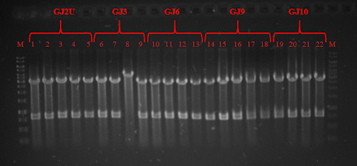
 Fig. 3 Restriction analysis of GJx constructs
 Fig. 4 Colony PCR of RNA thermometers in pSB1C3 plasmid.
 Fig. 5 expression at 24 ˚C. On the right you can see GFP expression without RNA thermometer.
 Fig. 6 GFP expression at 30 ˚C. On the right you can see GFP expression without RNA thermometer.
 Fig. 7 GFP expression in 37 ˚C. On the right you can see GFP expression without RNA thermometer.
 Fig. 8 Associational scheme of thermoswitches’ action in the SynDrop system. Not locking the concomitant translation of our target protein and BamA results in target protein aggregation due to insufficient membrane insertion and assembling potential of BamA.
 Fig. 9 Associational scheme of thermoswitches’ action in the SynDrop system. Locking up translation gives time for proper folding and insertion of BamA and prevents undesirable aggregation of target membrane proteins.


 Fig. 1 Simplified structure of scFv Antibody
 Fig. 2 Scheme of scFv_antiVLY and VLY interaction. Left- scFv_antiVLY binds to VLY, erythrocytes stay intact, Right- scFv_antiVLY does not bind and VLY lyse erythrocytes.
 Fig. 3 SDS-PAGE of scFv. GFP is used as positive control, C- chaperone DnaK.
 Fig. 4 Percentage of erythrocyte lysis at different +/-scFv dilutions.
 Fig. 5 A- MstX-scFv_antiVLY expression in Escherichia coli. B- scFv_antiVLY and MstX-scFv_antiVLY expression in cell-free system.
 Fig. 6 Fig 6. Percentage of erythrocyte lysis at different scFv/MstX-scFv dilutions.