Team:HK HCY LFC/Results
Results
In order to test for the feasibility of the DNA nanostructure, two parts of experiment were performed:
1. Test for the interaction between each single strand DNA oligos
2. Peroxidase activity assay
3. Cloning (Collaborated with HKU team)
First, the concentration of each single strand DNA oligos were compared to ensure that same
concentration of DNA oligos were used for assembly of the nanostructure. Each DNA oligos were
loaded into a 12 % polyacrylamide gel and the gel was post-stained using SYBR safe. The result is
shown in figure 1. It is observed that oligo 2 and 3 have a lower band intensity than that of oligo 1.
SYBR safe preferentially binds to double strand DNA (dsDNA) than single strand DNA (ssDNA) since
it is a intercalating dye. [1] Thus it is deduced that the difference in band intensity of the band is
mainly due to the length and conformation of the oligo rather than the concentration of the oligo.
Since oligo 1 has a larger size (62 bases) than oligo 2 (36 bases) and oligo 3 (39 bases), oligo 1 has
a larger secondary structure than oligo 2 and 3. Therefore, it has a higher chance of intercalating by
the dye.

Figure 4: Comparison of the concentration of the three DNA oligos using 12 % polyacrylamide gel
electrophoresis. Table on the right showing the content of each well.
After the comparison of the concentrations of each DNA oligo, the interaction of each DNA strand were tested and the DNA nanostructure was assembled using a thermocycler. The three DNA oligos were put together at 95oC for 5 min and cool down to 25oC with 0.5oC drop every 30 seconds. After that, the DNA solutions were loaded into a 12 % polyacrylamide gel.
The result is shown in figure 5. First, no excess banding of oligo 1,2 or 3 were shown in lane 5 to 10, so the concentration of each oligos are the same.
Second, Lane 5 to 7 shows the interactions between the two of the three DNA oligos. Oligo 1 binds with oligo 2 and 3 as predicted in lane 5 and 6 respectively. Oligo 2 and 3 will not bind to each other in our design but in the gel, it appears as one band in lane 7. It is deduced that the length of oligo 2 and 3 are too close, only 3 bases different, so the gel cannot separate the two oligos effectively.
Furthermore, it is observed that on lane 8, the three strands have interacted which result in a larger
banding size than the single strand (lane 2 to 4) and the interaction between two DNA strands (lane 5
to 7).Besides, when three strands were put together with the input RNA and DNA oligos, a band with
the largest size was observed in lane 9 and 10 respectively, which indicate there is binding between
the assembled DNA nanostructure and the input RNA and DNA oligo respectively. Therefore, it is
speculated that the three DNA oligos assembled to form the DNA nanotweezers and bind with the
input RNA oligo as predicted. Next, peroxidase activity essay is needed to further confirm the
formation of G-quadruplex upon their binding.

Figure 5: Validation of the assembly of DNA nanostructure and the binding between the DNA
nanostructure and the target oligos using 12 % polyacrylamide gel electrophoresis.
According to the design of our DNA nanostructure, G-quadruplex will be formed once it binds with the input RNA oligo. It is known that G-quadruplex will bind with hemin and possess a peroxidase activity. Thus, the formation of G-quadruplex can be confirmed by measuring the peroxidase activity. 2,2'-azino-bis(3-ethylbenzthiazoline-6-sulphonic acid) (ATBS), which is a colorimetric agents and usually used as a substrate with hydrogen peroxide for a peroxidase enzyme.[2,3] The Hemin/G-Quadruplex Dnazyme acts as a catalase to speed up the reaction between hydrogen peroxide (H2O2) and ABTS in which H2O2is reduced into H2O and ABTS is oxidized to form a green product which can be measured at 415 nm.
The DNA nanostructure, RNA oligos and hemin were first added into a microcentrifuge tube covered with aluminum foil. The mixture was incubated for 20 minutes to allow the formation of G-quadruplex and binding of hemin to the G-quadruplex formed. After that, H2O2and ABTS were added. The absorbance of the mixture which indicate the reaction rate between H2O2and ABTS2- was then measured in a 96-well plate using a spectrometer.

The mean absorbance at 415 nm for DNA nanostructure and target RNA oligo (0.1457) is
significantly higher than the other controls (DNA nanostructure: 0.073; target RNA oligo: 0.09;
DNA nanostructure and random RNA oligo: 0.079) with a p < 0.05 using One-way ANOVA
analysis.
Figure 6: Comparison of absorbance in different control setups.
In figure 6, the target RNA and the DNA nanostructure gave a relative low absorbance on its own when comparing the one with DNA nanostructure and input RNA put together. Also, when the DNA nanostructure was put together with a random RNA oligos, the absorbance is lower comparing with our target RNA oligo, it shows that the DNA nanostructure is specific to the target miRNA.
Furthermore, an experiment with different concentration of target RNA oligo was conducted. The concentration of our nanomachines will remain constant while the concentration of RNA oligo ranged from 0nM to 200nM. The absorbance of the mixture increases steadily with the increase of concentration of RNA oligos, they are positively related. Highest absorbance can be found at 200nM of RNA oligo while lowest absorbance can be found at 0nM of RNA oligo (figure 7).

Using the linear regression analysis, the absorbance at 415 nm under different concentration (nM) of input RNA oligo is shown above with R2 = 0.984 and p < 0.05.
Figure 7: The relationship between the concentration of RNA oligo and the absorbance at 415 nm. The concentration of input RNA oligo is directly proportional to the absorbance.
From the above graph, the regression line obtained is y=0.0009x+0.1298 (R2=0.9739). We can calculate the limit of detection (LOD) which indicates the smallest concentration of RNA oligos that can be reliably measured by an above procedure [4].
The LOD is calculated as follows:
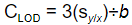
CLOD is the concentration LOD,
sylx : standard error of regression (SSE), and
b : slope of regression line.
The SSE obtained is 0.011212, the slope is 0.0023, thus the LOD is calculated as
3.3(0.011212) ÷ 0.0023 = 14.62 nM
As a result, the lowest concentration of RNA oligos which can be detected by our DNA nanostructure is 14.62 nM.
Three basic parts were submitted to the Registry. Each part code for one single strand DNA which make up the DNA nanostructure. Details of each parts is shown below.

Polymerase chain reaction (PCR) was conducted for the confirmation of plasmid. Primers are designed to amplify part of the DNA insert. The result is shown in figure 8.
There are two batches of plasmids (lane 2-4 and lane 5-7) shown. The brightest bands in lane 5 to 7 (surrounded by a red box) are believed to be the part of the DNA insert amplified. However, the specificity of the primers are not good, multiple bandings are shown. Thus, further validation is needed.

Figure 8: Validation of the plasmids with the DNA inserts amplified by PCR. The band surrounded in red box is the part of the DNA insert amplified.
From the experimental result, the three DNA oligos designed was successfully assembled to form a nanotweezers. It binds with the target RNA oligo to form a G-quadruplex. The peroxidase assay confirms that the formation of G-quadruplex is directly proportional to the concentration of the target mRNA 25. However, for the cloning part, further validation of the plasmid is needed.
References
[1] Haines, A. M., Tobe, S. S., Kobus, H. J., & Linacre, A. (2015). Properties of nucleic acid staining dyes used in gel electrophoresis. Electrophoresis, 36(6), 941-944.
[2] Wu, Y., Zou, L., Lei, S., Yu, Q., & Ye, B. (2017). Highly sensitive electrochemical thrombin aptasensor based on peptide-enhanced electrocatalysis of hemin/G-quadruplex and nanocomposite as nanocarrier. Biosensors and Bioelectronics, 97, 317-324.
[3] Shiu, S. C. C., Cheung, Y. W., Dirkzwager, R. M., Liang, S., Kinghorn, A. B., Fraser, L. A., ... & Tanner, J. A. (2017). Aptamer‐Mediated Protein Molecular Recognition Driving a DNA Tweezer Nanomachine. Advanced Biosystems, 1(1-2), 1600006.
[4] Shrivastava, A., & Gupta, V. B. (2011). Methods for the determination of limit of detection and limit of quantitation of the analytical methods. Chronicles of Young Scientists, 2(1), 21.


© IGEM HCYxLFC 2018 - All Rights Reserved.