Difference between revisions of "Team:BJRS China/Demonstrate"
| Line 126: | Line 126: | ||
<br/> | <br/> | ||
<h2>Surface display system</h2> | <h2>Surface display system</h2> | ||
| − | <p><font size="4">  We demonstrated the working effect of INP, intimin and autotransporter surface display system with the cell lysis experiment and microscope examination. The result of cell lysis experiment showed that after cell lysis followed by centrifuge, both the precipitation and supernatant shows relative strong fluorescence signal in intracellular expressing fluorescent protein cells, while the signal was stronger in supernatant than in precipitation in surface | + | <p><font size="4">  We demonstrated the working effect of INP, intimin and autotransporter surface display system with the cell lysis experiment and microscope examination. The result of cell lysis experiment showed that after cell lysis followed by centrifuge, both the precipitation and supernatant shows relative strong fluorescence signal in intracellular expressing fluorescent protein cells, while the signal was stronger in supernatant than in precipitation in surface displaying fluorescent protein cells, suggesting that the surface displayed fluorescent protein were anchored to the outer membrane and being precipitated with the cell fragment, while the intracellular expressed fluorescent protein were solved in the supernatant(fig.2).</font></p> |
<figure class="text-center"> | <figure class="text-center"> | ||
<img src="https://static.igem.org/mediawiki/2018/b/bd/T--BJRS_China--demo1.jpg" width="60%" > | <img src="https://static.igem.org/mediawiki/2018/b/bd/T--BJRS_China--demo1.jpg" width="60%" > | ||
| Line 133: | Line 133: | ||
</figure> | </figure> | ||
<br/> | <br/> | ||
| + | <p><font size="4">  From the microscopy examination(fig.3) we can see that the intracellular expression of fluorescent protein displayed the rode-shape of <i>E.coli</i>, while the signal of surface displayed fluorescent protein showed the dotted pattern around the cells(as the arrows indicated), which suggests that the fluorescent protein was gathered and distributed at the surface of <I>E.coli</i>. | ||
| + | </font></p> | ||
| + | |||
| + | </div> | ||
</html> | </html> | ||
Revision as of 17:22, 16 October 2018
Demonstrate
Overview
The aim of our project is to increase the oxygen carrying capacity of engineering bacteria. Previous work have shown that the expression of bacterial Vitreoscilla hemoglobin (VHb) can help bacteria utilize oxygen more efficiently[1]. However, the bacterial cell membrane makes efficient hemoglobin-oxygen contact a challenge. Based on this, our team designed a VHb surface display system to express VHb on the outermost shell of the bacteria to raise the hemoglobin-oxygen contacting efficiency. We named this system OxygenMAX system.
Firstly, we conducted a model to predict the effect of VHb expressing and the gathering of oxygen by VHb on the growth rate of bacteria, which came out that the surface display of VHb can help the bacteria grow faster in theory.
We found three kind of surface display system respectively are INP, intimin and autotransporter. We linked fluorescent protein to them separately to check if they work by the cell lysis experiment and the microscope examination, and our results suggested that all the three surface display system work.
Finally we conducted the growth curve measurement experiment to see if our oxygenMAX system worked. From the result we can see that the VHb surface displayed bacteria grew faster than the wildtype bacteria on the the exponential phase.
Therefore, we believe that our project can reach the medal requirement of demonstration. For more details please see our modeling page and results page.
Modeling
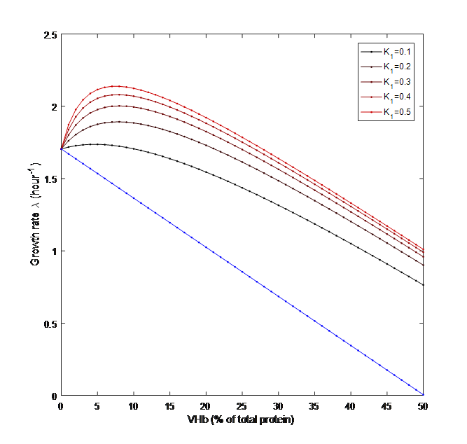
We built a model based on the influence of oxygen concentration and protein expression on the growth rate of bacteria. We hypothesized that the VHb can influence the oxygen concentration in a liner relation with a coefficient K1 and that surface display of VHb can increase the value of K1. Finally we got the prediction of the effect of the VHb percentage and K1 on the growth rate of bacreria.(fig.1)
Surface display system
We demonstrated the working effect of INP, intimin and autotransporter surface display system with the cell lysis experiment and microscope examination. The result of cell lysis experiment showed that after cell lysis followed by centrifuge, both the precipitation and supernatant shows relative strong fluorescence signal in intracellular expressing fluorescent protein cells, while the signal was stronger in supernatant than in precipitation in surface displaying fluorescent protein cells, suggesting that the surface displayed fluorescent protein were anchored to the outer membrane and being precipitated with the cell fragment, while the intracellular expressed fluorescent protein were solved in the supernatant(fig.2).

From the microscopy examination(fig.3) we can see that the intracellular expression of fluorescent protein displayed the rode-shape of E.coli, while the signal of surface displayed fluorescent protein showed the dotted pattern around the cells(as the arrows indicated), which suggests that the fluorescent protein was gathered and distributed at the surface of E.coli.